
Twice a year the Crane Trust science team collects data on our Bison’s parasitic loads, once in May (start of growing season) and once in August (end of growing season). This bi-yearly sampling helps us keep a pulse on which individuals have what parasites, how large their parasitic loads are, and if the parasites are affecting the bison’s health. We count the exact amount of Strongyle, Moniezia, Nematodirus, Trichurus, and Coccidia. In the same order; these parasites are more commonly identified as bloodworms, tapeworms, roundworms, whipworms, and obligate, intracellular protists.
Why?
Parasites typically have negative effects on their hosts. Because of this, we keep an eye on our bisons’ parasitic loads. However, through previous monitoring, we have not seen health declines in bison who have more parasites. This gave us reason to stop giving anti-parasite medicines (spray-on dermal and oral dewormers), allowing us to further care for our bison in a natural, more hands-off approach as it seems the bison gain immunity as they age. Intervention is only needed in the occurrence of weight reduction, reproductive difficulties, and possible death; which we haven't encountered yet. However, we still have questions regarding our bison and parasites This gives us reason to continue monitoring parasites 1.) Do cows with calves have more or fewer parasites? 2.) What do a calf’s parasite loads look like in comparison to their mothers? Do they correlate?
Sample Collecting
Before the bison patty party can start someone has to collect bison feces. Josh Wiese, Range Manager, and Mallory Beckman, Range Technician, usually collect samples. When collecting, they wait and watch for individuals to relieve themselves, drive up to the blast zone, and scoop a sample into a plastic bag with a single-use plastic spoon. All of this is done with safety in mind. They keep ample distance between the bison and themselves and only pick up samples in safe locations. These plastic bags are labeled with the bison's identification number. If you would like to know more about how we complete the process of extracting these parasites from our bison’s feces, I have included our step-by-step instructions below.
Step-By-Step Instructions: Bison Fecal Flotation Procedure
1. Squeeze the bag containing the fecal sample until the contents are well mixed.
2. Weigh 5g of fecal sample out into a clean weighing container (cup).
3. Add 20 ml of deionized water to the cup and mix thoroughly, breaking the fecal matter apart while mixing.
4. Filter out the solid fecal matter using a tea strainer and tongue depressor to collect as much of the liquid portion of the mixture as possible into a new cup to help separate the parasite eggs from the fecal matter.
5. Divide the liquid evenly into two 15 ml capped conical tubes and label each with the bison tag number corresponding to the fecal sample. Filling each conical tube with the same amount of liquid helps keep the centrifuge balanced.
6. Centrifuge the sample (properly balanced) at 650 x g (2500 rpm) for 10 min. and allow the centrifuge to stop without braking. This forces all the suspended solids in the liquid to the bottom of the tube.
7. Remove the conical tubes and discard the liquid portion(supernatant) and add 11 ml of Sheather's flotation solution to each tube. Resuspend the fecal pellet by mixing each tube with a stir-rod (wooden portion of a long cotton swab) and completely breaking the pellet apart. Continue filling each tube to the same amount and put on the cap.
8. Return the tubes to the centrifuge (balanced) at 650 × g (2500 rpm) for 10 min., allowing the centrifuge to stop without braking. The sheather’s solution is heavier than the parasite eggs. Centrifuging them forces the eggs to the top.
9. Remove the tubes from the centrifuge after spinning and place them into a test tube rack.
10. Slowly fill each tube with Sheather's flotation solution to form a slight bubble of liquid on the top of the tube.
11. Place a cover slip on top of each tube and allow it to sit for 30 min. Since the eggs are at the top of the tube this allows them to collect on the cover slip.
12. Carefully lift the coverslips off and place them side-by-side on a clean microscope slide.
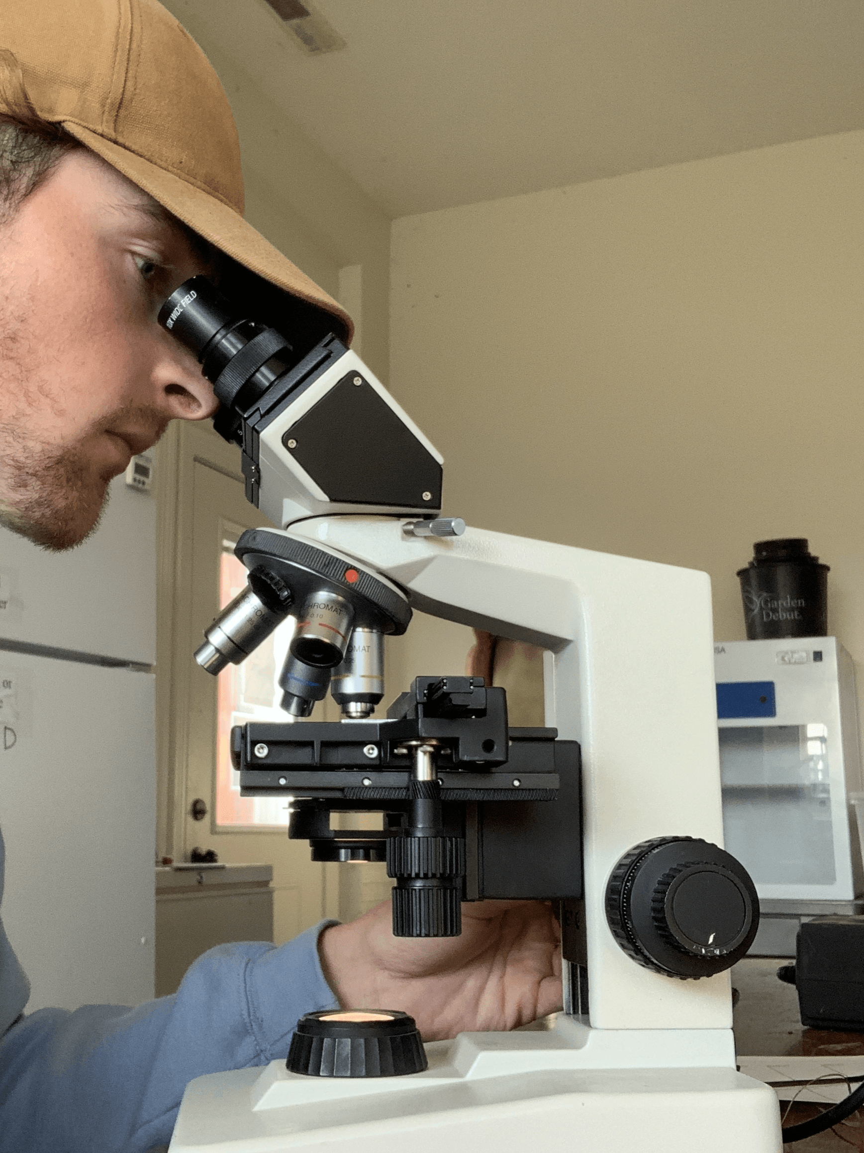
13. Read both coverslips under 10x magnification and count each parasite egg type separately.
14. Divide the sum of each egg type by 5 to provide the number of parasite eggs per gram of feces.
15. Record the sampling date, tag number, and parasite eggs per gram on a datasheet and add them to the bison parasite database.
Parasite Life Cycle
So now that you understand how we study bison patties, let's explore the life cycle of parasites in general. Parasites usually have one or two or more hosts. When a parasite takes advantage of two or more hosts, the hosts not needed for reproduction aid in the transportation and development of the parasite. Once a parasite has made it to the final host, it can travel to the organ(s) or tissue(s) that it has adapted to use and release eggs or oocysts, which are protozoan zygotes. These are released through excrement in our Bison and then the cycle will continue.
How Bison Acquire Digestive Parasites
Our bison’s digestive system parasites most commonly have only one host, the bison. They will defecate these eggs and oocysts. After their release, the next bison will ingest some parasites while foraging for their preferred plants in the future. When two hosts are present, mites or other small arthropods help transport parasites to our bison.
Our Mission with Bison
At the Crane Trust, we aim to be better stewards of our land. Our bison help us do that by providing different grazing and disturbance methods to our collection of management techniques. Our philosophy is that we need to be willing to change our practices when evidence suggests to continually better maintain our land. We will never have a perfect management style, but we are always aiming to improve it and work through problems holistically.
My Experience
Microscope work is something I enjoy despite some individual’s parasites taking a while to count, constantly refocusing, and having to handle feces. When you look into a microscope you enter another world. A world of the small things you will never get to see with your naked eye. This creates a sense of wonder for what is hiding from you in every other place you haven't been able to look.
See you in the next blog,
Matt Urbanski, Saunders Conservation Fellow
murbanski@cranetrust.org